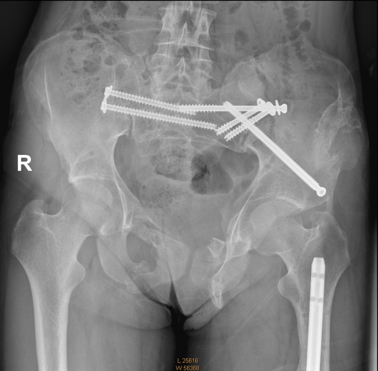
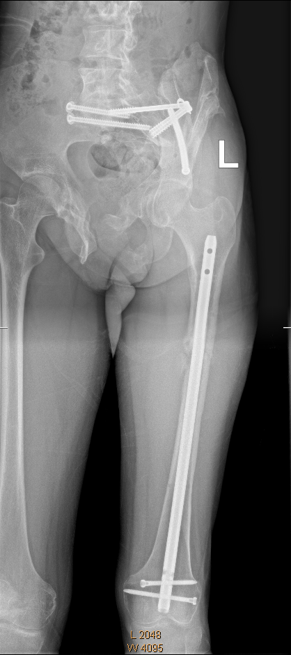
Department: Trauma / orthopedic
Patient: female – age: 22Y
Dr. Amal Khoury
Services: Segmentation, surgery planning
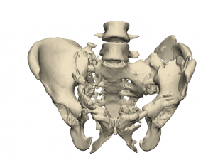
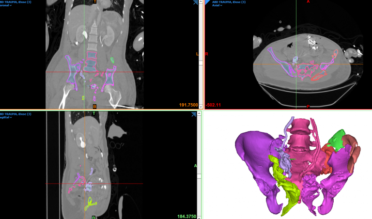
This case began with the CT imaging we got from Dr. Khoury and started to make a 3D imaging out of the dicom.
Our workflow goes as following: we extract one 3D part, including all the bony areas which are relatively bright colored, in all range of black & white image.
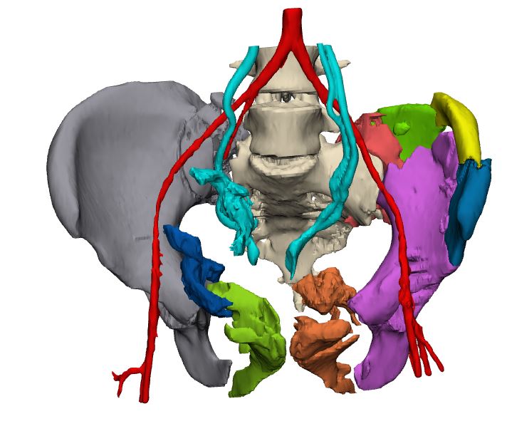
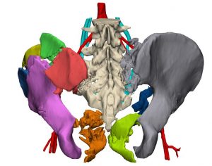
Then, we split this one-part anatomy into fragments, each fragment to be colored on different color, to differ between them. This following case was relatively drastic by its fragments due to the drastic trauma cause, so, we had relatively big amount of fragments.
After splitting is done, the doctor and the medical planner move the different fragments, like a 3D puzzle, to fit each other and together would form the original anatomy as much as possible.
