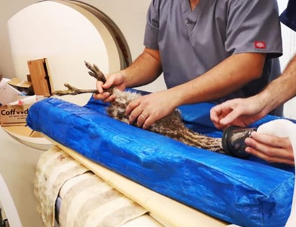
In the case of an ostrich presented with a limb deformity, we utilized CT imaging to 3D build the anatomical anomaly. With this data, we planned a 3D-printed model of the ostrich’s limb, providing the veterinarian with a tangible comprehention of the unique condition. This innovative approach facilitated a deeper understanding of the deformity’s complexity, affecting the veterinary team to develop a tailored treatment plan to the ostrich’s specific needs.