Department: spine (Pediatric) Patient: male, age: 2Y Dr. Amit Sigal. Services: Segmentation, surgery planning, anatomy model, PSI
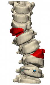
The most frequent cause of congenital scoliosis is a hemivertebrae. This not-fully-developed bony tissue, causing the spine to be curvy. The patient is not fully symmetric and can be with one shoulder above another, one hip above the other etc.
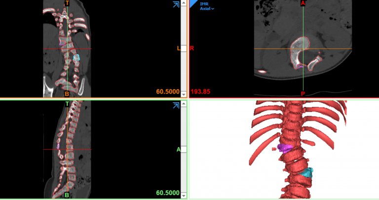
When we get the CT from Dr. Sigal we transform it into virtual 3D and with the doctor, we insert screws inside the vertebrae’s pedicels. Screws must not penetrate the vertebrae volume and must not touch the veins adjacent to the vertebrae. This may cause unwanted results. The virtual platform allows us to see the vertebrae transparently and so, simplify the surgery plan and insert the screws in a much easier way.
Once
the screws are located, we match K-wires to their location. The K-wire
functions a preparation for the screw drilling.
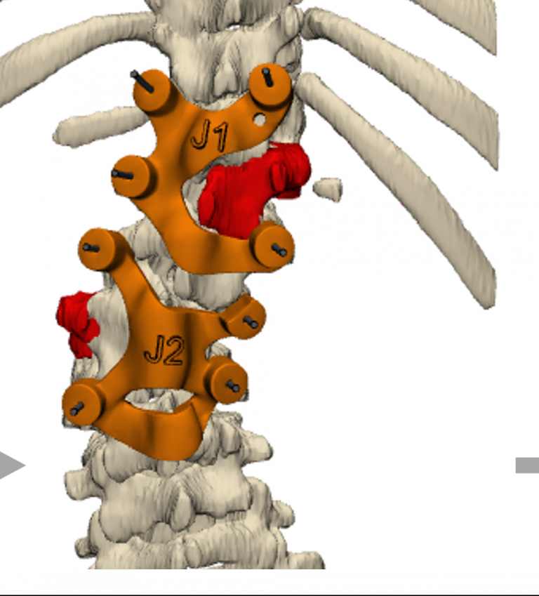
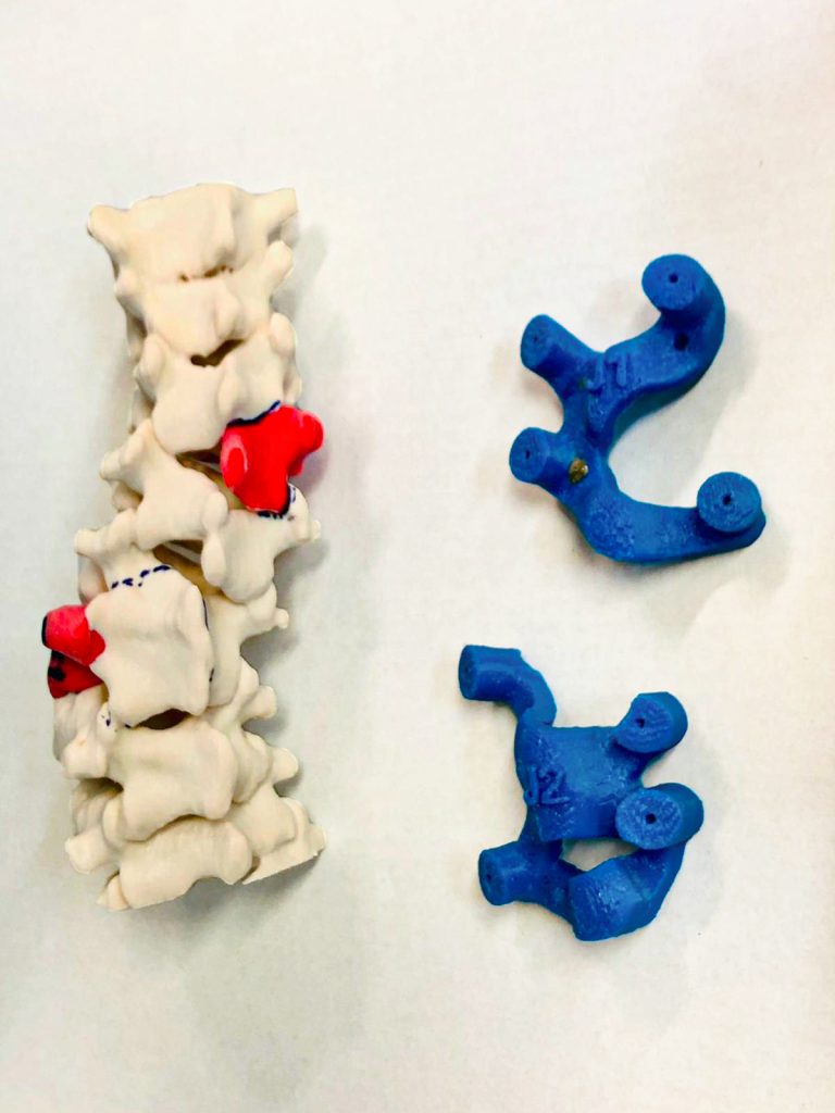
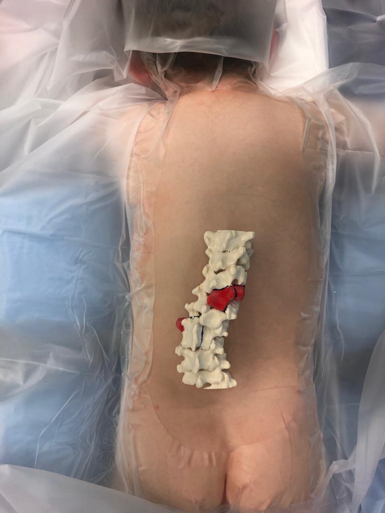
The jig, supplied to operation, is sterile and located on a specific place on
the patient’s anatomy, showing the doctor how to apply the virtual planning.
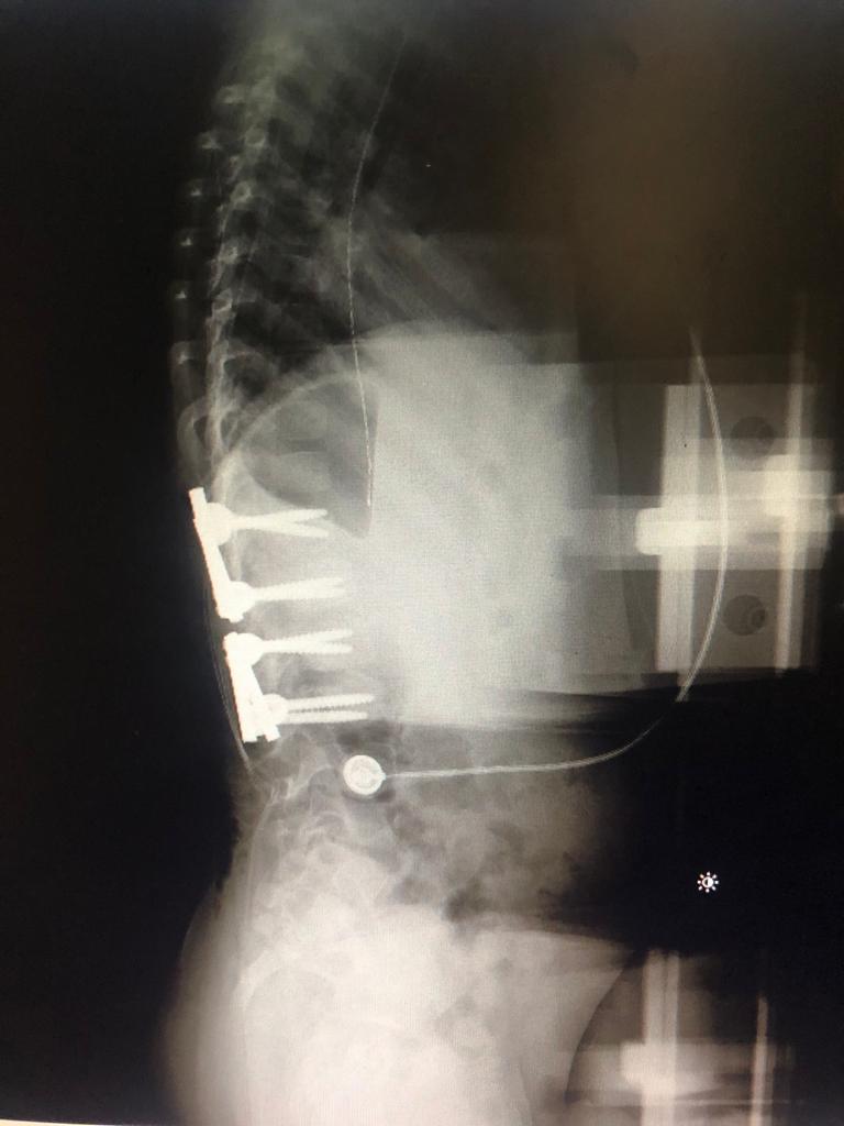
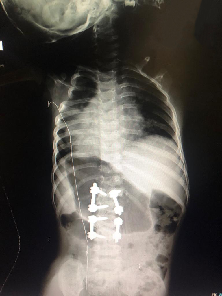
The patient’s spine is fixed by screws and guiding rods, keeping it straight
and healthy