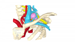
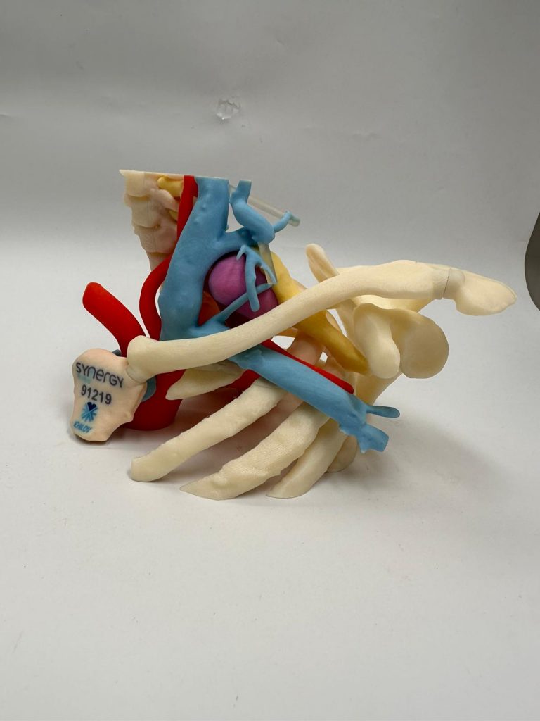
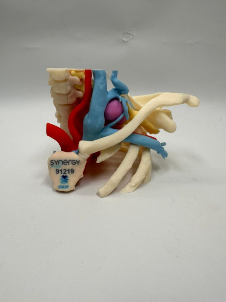
A woman was diagnosed with a tumor situated behind her clavicle bone, in close proximity to a major blood vessel, the inferior vena cava (IVC). Upon reviewing her medical imaging, the doctors determined that utilizing a 3D model would provide them with a more detailed and comprehensive understanding of her condition. This model would assist them in strategizing the best approach for treatment.
To achieve this, the medical team virtually designed a precise 3D representation of the affected area. The model was then 3D printed, incorporating multiple colors to clearly differentiate and distinguish between various anatomical structures. This advanced visualization tool enabled the doctors to plan and execute a more effective and targeted treatment plan for the patient.