This is a neurosurgery case that we took to the next step using our cutting-edge 3D technology.It usually comprises of a two-part surgery. A craniectomy and a cranioplasty.
As the human brain is protected by the skull, it is also a an incompressible ‘closed box’. In traumatic brain injury or stroke aside from the event itself, and secondary brain damage can be caused by the increased pressure inside the ‘closed box’ (skull), whether from the bleeding in traumatic brain injury or from the edema and swelling of the brain. A decompressive craniectomy surgery can prevent the secondary neurological damages.
In this case, in cooperation with the surgeon, we created a
customized implant. We used the CT taken after the craniectomy to supply the patient with a perfectly fitting piece for the skull.
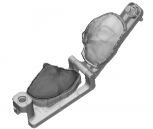
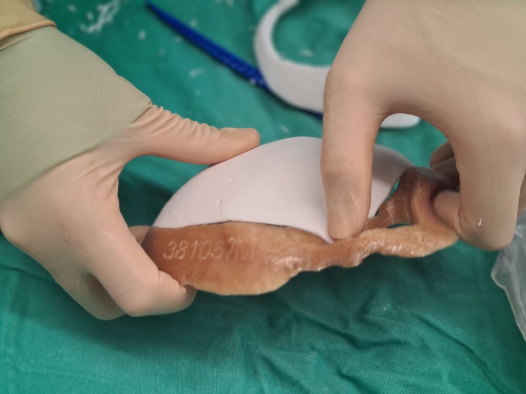
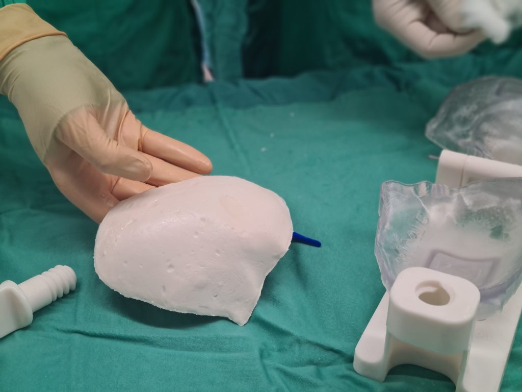
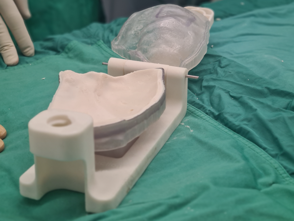
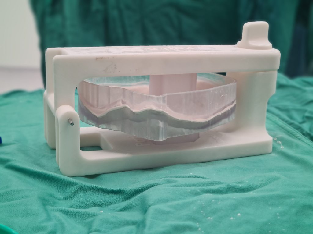
Once the implant’s shape is approved, a 3D printed patient-specific kit is being printed. It consists of molds and clamp, both of which are sterilized prior to surgery. During the surgery, the surgeon pours bone cement into the 3D printed customized mold. This cement piece can be tested on a sterile 3D printed anatomical model, prior to using it on the patient. The sterilization serves to reduce the risk of secondary infection.
Using our technology, surgeons can reduce surgery time. It also resulted in a perfectly fitting and symmetrical implant for the patient.